Home
![]()
Deep-Learning Inferred Multiplex Immunofluorescence for IHC Image Quantification
Read Link | Cloud Deployment | Docker | Report Bug
Reporting biomarkers assessed by routine immunohistochemical (IHC) staining of tissue is broadly used in diagnostic pathology laboratories for patient care. To date, clinical reporting is predominantly qualitative or semi-quantitative. By creating a multitask deep learning framework referred to as DeepLIIF, we present a single-step solution to stain deconvolution/separation, cell segmentation, and quantitative single-cell IHC scoring. Leveraging a unique de novo dataset of co-registered IHC and multiplex immunofluorescence (mpIF) staining of the same slides, we segment and translate low-cost and prevalent IHC slides to more expensive-yet-informative mpIF images, while simultaneously providing the essential ground truth for the superimposed brightfield IHC channels. Moreover, a new nuclear-envelop stain, LAP2beta, with high (>95%) cell coverage is introduced to improve cell delineation/segmentation and protein expression quantification on IHC slides. By simultaneously translating input IHC images to clean/separated mpIF channels and performing cell segmentation/classification, we show that our model trained on clean IHC Ki67 data can generalize to more noisy and artifact-ridden images as well as other nuclear and non-nuclear markers such as CD3, CD8, BCL2, BCL6, MYC, MUM1, CD10, and TP53. We thoroughly evaluate our method on publicly available benchmark datasets as well as against pathologists' semi-quantitative scoring.
© This code is made available for non-commercial academic purposes.
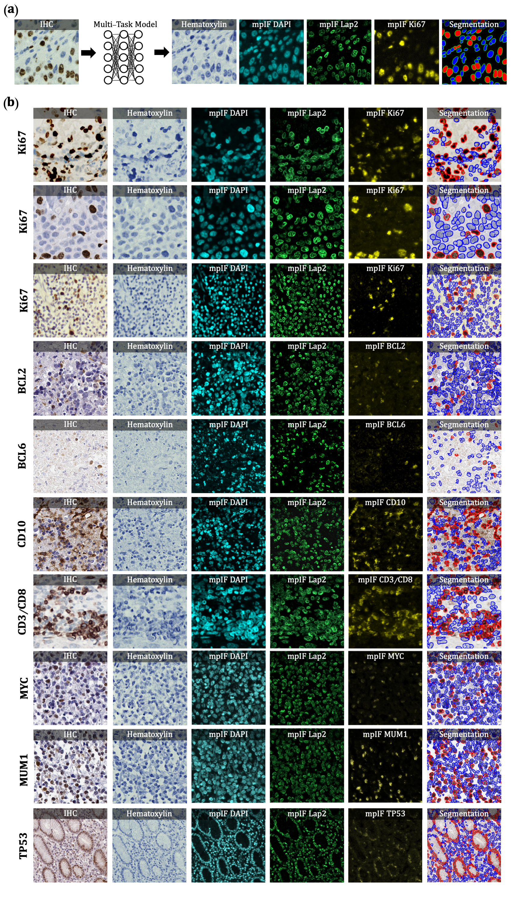 Overview of DeepLIIF pipeline and sample input IHCs (different
brown/DAB markers -- BCL2, BCL6, CD10, CD3/CD8, Ki67) with corresponding DeepLIIF-generated hematoxylin/mpIF modalities
and classified (positive (red) and negative (blue) cell) segmentation masks. (a) Overview of DeepLIIF. Given an IHC
input, our multitask deep learning framework simultaneously infers corresponding Hematoxylin channel, mpIF DAPI, mpIF
protein expression (Ki67, CD3, CD8, etc.), and the positive/negative protein cell segmentation, baking explainability
and interpretability into the model itself rather than relying on coarse activation/attention maps. In the segmentation
mask, the red cells denote cells with positive protein expression (brown/DAB cells in the input IHC), whereas blue cells
represent negative cells (blue cells in the input IHC). (b) Example DeepLIIF-generated hematoxylin/mpIF modalities and
segmentation masks for different IHC markers. DeepLIIF, trained on clean IHC Ki67 nuclear marker images, can generalize
to noisier as well as other IHC nuclear/cytoplasmic marker images.
Overview of DeepLIIF pipeline and sample input IHCs (different
brown/DAB markers -- BCL2, BCL6, CD10, CD3/CD8, Ki67) with corresponding DeepLIIF-generated hematoxylin/mpIF modalities
and classified (positive (red) and negative (blue) cell) segmentation masks. (a) Overview of DeepLIIF. Given an IHC
input, our multitask deep learning framework simultaneously infers corresponding Hematoxylin channel, mpIF DAPI, mpIF
protein expression (Ki67, CD3, CD8, etc.), and the positive/negative protein cell segmentation, baking explainability
and interpretability into the model itself rather than relying on coarse activation/attention maps. In the segmentation
mask, the red cells denote cells with positive protein expression (brown/DAB cells in the input IHC), whereas blue cells
represent negative cells (blue cells in the input IHC). (b) Example DeepLIIF-generated hematoxylin/mpIF modalities and
segmentation masks for different IHC markers. DeepLIIF, trained on clean IHC Ki67 nuclear marker images, can generalize
to noisier as well as other IHC nuclear/cytoplasmic marker images.